This project applies two novel technologies to quantify cancer cell phenotypes using cells from immortalized cell lines and cells disaggregated from human biopsies. The first novel technology is a hermetically-sealed microenvironmental chamber that allows respiration rate and ion flux measurements. The second technology is an optical CT scanner for 3D cell imaging that facilitates extraction of nuclear morphometric features.

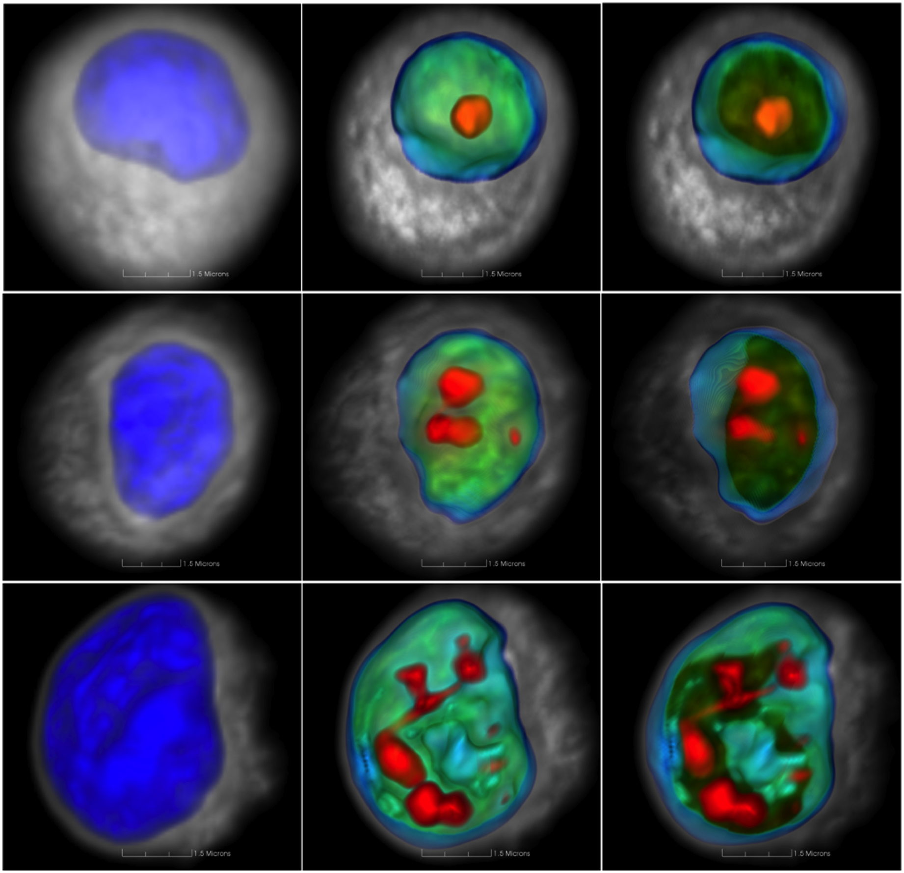
Volume renderings of EPC (top), CP-C (middle) and CP-D (bottom) esophageal epithelial cells. Left images show the nuclear surface, middle images illustrate the nuclear interior, and right images depict a slab through the volume.

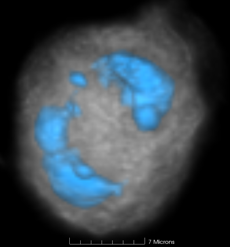
Pseudo-color volume rendering of MDA-MB-231 cell imaged using cell CT. The image illustrates what appear to be multiple nuclei (depicted in blue) within and intact cell membrane. The distorted shape of the nuclei are notable. The cytoplasm is shown in grey.
Atomic Force Microscopy: ‘hands-on’ – April 15th to 16th 2010, ASU Tempe and Agilent


